
1-8 µm
Voxel resolution achievable
25 mm
Max sample size
20 mp
Single shot capture
2 Channels
E.g. HREM + Lac-Z
WHOS IT DESIGNED FOR
The right system for your lab?
The OHREM Micro is built for individual research labs that want to do HREM imaging in-house with a straightforward workflow. It is not a multi-user platform focused, cost-effective tool for groups with a clear application in mind.
The Micro is a good fit if you…
-
Are an individual lab or small group doing routine HREM imaging
-
Work primarily with standard HREM morphology, one channel for most runs
-
Want to add a second channel for LacZ reporter imaging alongside standard HREM
-
Routinely image samples in the 1–15mm range as your main use case
-
Want a lower-cost entry point to in-house HREM with room to upgrade later
-
Don't need automated multi-sample or scanning workflows from day one
Consider the Ultra instead if you…
-
Run a core facility or high-throughput imaging programme
-
Need true fluorescence imaging (GFP, mCherry, multi-channel beyond LacZ)
-
Want to image multiple samples in one block with higher resolution
-
Need automated XY scanning with encoded stage control as standard
-
Are working with samples larger than 25mm or up to 70mm
-
Want to experiment with structured illumination for SBF from the start
OVERVIEW
Simplified HREM for Research
The Optical HREM Micro is a smaller form factor, benchtop system that brings high-resolution episcopic microscopy into the individual lab without the overhead of a large multi-user platform. It produces inherently aligned 3D image stacks at voxel resolutions down to 1 micron, using a fixed optical configuration and a straightforward acquisition workflow through OHREM Acquire software.
The Micro is built around a fixed optical platform, there is no automated encoded XY stage as standard, which keeps the system simpler and more affordable. Manual X and Y adjustments allow sample centring, but if your work requires automated scanning or multi-sample throughput, the OHREM Ultra is the appropriate choice.
Each system is hand-assembled and tested to order by the Indigo Scientific engineering team, shipped with a pre-configured PC, starter consumables and on-site training. A 1-year warranty with parts and team access is included as standard.
Dense, uncleared samples
Images resin-embedded opaque tissue directly, no optical clearing required.
Simple workflow
Straightforward acquisition through OHREM Acquire with minimal setup per run.
Inherently aligned output
No registration step. Stacks export ready for immediate 3D reconstruction.
Built in-house
Hand-assembled and tested to order by the team that designs the systems.

Optical HREM Micro — imaging stage with resin block illuminated
MICRO IMAGING
Single-shot imaging as standard

Fixed
Optics platform as standard, no automated encoded XY stage
Add-on
XY scanning available as an upgrade at order or retrofitted late
Auto
When fitted, stitching is automatic through OHREM Acquire software
A note on fluorescence channels
The Micro supports up to 2 illumination channels, standard HREM morphology plus a second channel suitable for LacZ reporter imaging. This is not a full fluorescence platform. If your work requires GFP, mCherry or multi-channel fluorescence imaging, the OHREM Ultra is the correct system, with up to 8 channels and adaptions to improve fluorescence imaging.

HREM Micro output — mouse embryo cross-section · 20 MP monochrome
APPLICATIONS
What the OHREM Micro images
The Micro is closely related to our original Optical HREM system used extensively in cardiovascular and developmental biology research. Its strength is consistent, cost-effective routine morphological imaging.
Cardiovascular Research
Closely related to the original HREM system used extensively for cardiovascular research. Visualise 3D cardiac structures, study congenital heart defects and phenotype mutant mouse lines from E9.5 onwards.
Developmental Biology
Whole mouse embryo imaging from E9.5 through to E18.5. Full 3D reconstruction of embryo morphology, tracking developmental abnormalities and quantifying organ formation and spatial relationships.
Plant Sciences & Others
3D imaging of seed structures, root development, vascular bundles and meristems. Also zebrafish larvae, Drosophila and other model organisms in the appropriate size range.
Small Animal Organs
Kidney, liver, placenta and lung from under 1mm to 20mm. Measure volume, surface area and branching networks with consistent micron-scale section thickness.
TECHNICAL SPECIFICATIONS
OHREM Micro specificationn
Standard Micro configuration. Many parameters can be adapted at order, contact us to discuss.
Optics & imaging
Optical platform
Samples up to 20 × 20 × 25 mm
Variable zoom optic
Included as standard
Image capture
20 megapixel monochrome
Section thickness
1–8 μm (defines axial resolution)
Output formats
TIFF, TIF, BMP, PNG, JPEG, JPG
Included with purchase
Software
OHREM Acquire — no licence fee
PC & setup
Pre-configured, drivers installed
Warranty
1 year — parts + team access
Training
On-site 1–2 days at installation
Consumables
1 split mould + 20 chucks
Stage & mechanics
XY stage
Fixed — manual centring only*
Encoder
Yes — exact section thickness
Sample extraction
Included — prevents debris on block
Compatible blades
60mm and 80mm (single use)
Multi-sample
Not standard — single sample per run
Available add-ons
XY scanning stage
Small motorised XY — tile scanning
Second channel (e.g LacZ)
Filter wheel/slider for 2nd chann
Wavelength focus adj.
Focus correction across channels
Motorised focus
Desktop controller, fine adjustment
Alternative imaging
Higher resolution / sensitivity etc.
*X and Y adjustments allow sample centring but there is no automated encoded stage. For precise XY control, the add-on stage is available — however if automated scanning is core to your workflow, see the OHREM Ultra.
† For routine imaging of samples larger than 15mm the Ultra is the advised system.
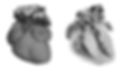
HREM Micro 3D Mouse Heart
OHREM Micro — FAQ
What sample sizes does the Micro support?
Samples up to 20 × 20 × 25mm. There is effectively no lower limit, the system handles specimens below 1mm. For samples larger than 25mm, the OHREM Ultra is the correct system, supporting up to 70 × 70 × 35mm with the Z height increase add-on available for 50mm.
Can the Micro do fluorescence imaging?
The Micro supports up to 2 illumination channels the standard HREM channel plus a second channel suitable for LacZ reporter imaging. It is not designed as a fluorescence platform. For GFP, mCherry, multi-channel fluorescence or optical sectioning, the OHREM Ultra is the appropriate system. Fluorescence must be combined with a compatible MF-HREM protocol and additions to improve contrast.
Does the Micro come with an XY scanning stage?
No, the Micro has a fixed optical platform as standard with manual X and Y centring adjustments. An XY scanning stage add-on is available at purchase or as a retrofit, but if automated scanning is a core requirement from day one, the OHREM Ultra includes an encoded XY stage as standard.
Can I Image multiple samples in the same block?
The Micro is designed for single sample per run. Multiple samples can be embedded in one block, but without an automated XY stage, imaging more than one sample in a block requires the XY add-on. The OHREM Ultra is designed for multi-sample throughput with dedicated multi-well holders.
What blades does the Micro use?
The Micro is compatible with 60mm and 80mm tungsten carbide blades. These are single-use blades. The OHREM Ultra additionally supports 160mm reusable tungsten carbide blades for extended runs.
Can the Micro be upgraded after purchase?
Yes — the XY scanning stage, second fluorescence channel (LacZ), wavelength focus adjustment and motorised focus control can all be retrofitted. Contact us with your system details and we will advise on what is applicable. Note that the Micro cannot be upgraded to Ultra capability — the Ultra is a different platform.
What software is included and what analysis platform does it support?
OHREM Acquire is included — no licence fee, free updates. Free additional tools including Image Viewer, Stack Editor, Translation Tools and 3D Viewer are available from our software page. Output TIFF stacks are compatible with Fiji/ImageJ, Imaris, Dragonfly, 3D Slicer and Imalytics Preclinical.
Do you ship, install and service outside the UK?
Indigo Scientific serves worldwide, contact us for more information.
Indigo Scientific - HREM Team
Based in Hertfordshire, UK. Systems shipped and supported worldwide.