top of page
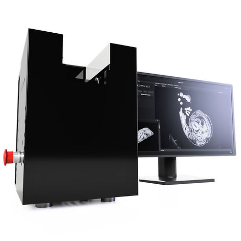
Optical High Resolution Episcopic Microscopy(HREM) System

High Resolution Episcopic Microscopy (HREM) is a 3D biomedical imaging technique comprising of 2D sections for structural analysis of samples.

What is High-Resolution Episcopic Microscopy(HREM)?

High-Resolution Episcopic Microscopy (HREM) is a 3D imaging technique for visualising samples ranging from 1-30mm. Resin mounted samples are stained, then sectioned repeatedly with the remaining block face imaged, creating high-resolution stacks at 1–8-micron voxel resolution.

HREM imaging technique offers some unique benefits:

  • Produces 1-8 micron complete voxel resolution over an entire volumetric dataset

  • Image dense tissue and bone at high contrast without variation

  • No sample clearing, distortion or interpolation

  • Capable of imaging up to 60mm samples

We offer two solutions for performing HREM, the Micro and Ultra. As well as other products.

Embryonic mouse heart imaged using optical hrem block face imaging system.
HREM Ultra Block Face Scanning

Advantages

High Resolution Episcopic Microscopy (HREM) as a 3D imaging technique of ex-vivo samples gives multiple benefits over other techniques such as resolution, cost and more.

Flexibility for a Range of Samples

High-Resolution Episcopic Microscopy(HREM) provides exceptional clarity and resolution, even when imaging denser tissues or tissues with varying opacities.


Unlike conventional techniques, eliminate the need for tissue clearing and interpolation, making the imaging process more efficient and convenient. HREM produces high-contrast results, enhancing the visualization of anatomical structures and details.

  • Capable of visualising samples up to 30mm in one capture or greater with scanning

  • Suitable for bone and denser tissue samples

  • High contrast images even in higher density areas

A Range of High Resolution Episcopic Microscopy (HREM) samples including 3D image of a zebrafish, 3D image of a mouse embryo, 3D image of a chicken brain, 3D image of a heart and a 3D image of barley. All captured using the OHREM instruments.

High Resolution for All Tissue and Sample Types

3D Biomedical imaging techniques have evolved significantly, with HREM standing out for its ability to deliver precise 3D tissue images of denser samples. High-Resolution Episcopic Microscopy (HREM) offers a variety of resolutions with scanning stages and a choice of optics. It can capture single shots or high-resolution scans at below 1 micron voxel size magnification with blending, no blocking.

With sections as thin as 1 µm, from our speciality sectioning system, HREM achieves voxel resolutions of 1-8 µm³, enabling the identification of fine details like individual nerves and blood vessels, surpassing other techniques.

  • 1-8 micron voxel size resolution

  • XY scanning stage integrated into the software available for larger samples

HREM 3D embryonic mouse cross section captured with HREM morphology imaging system

Easy Data with HREM

The use of 3D imaging in medical research is growing rapidly, with HREM providing accurate and detailed views of tissue samples. High-Resolution Episcopic Microscopy (HREM) data is readily applicable for both 2D and 3D analysis. Simple software packages like Fiji and Dragonfly can be used to generate impressive images by inverting the LUT (Lookup Table). 


HREM images can be manually or automatically segmented in the user-friendly 2-D image format, enabling precise measurements.

  • Data is stored as image files for easy manipulation (jpeg,tif and more)

  • Stored image sizes for easy workflow

  • No custom image analysis software required

High Resolution Episcopic Microscopy Applications

High-Resolution Episcopic Microscopy (HREM) is used to study a variety of biological samples, including mouse organs such as brains and hearts, whole mouse and zebrafish embryos, plant matter, drosophila, and more applications for 3D imaging.​​​

Optical HREM Systems

We offer two main Optical HREM system solutions, available worldwide, offering flexibility in budget and in capture. Each systems can achieve highly detailed 3D image stacks, while allowing the systems to be accessible to more.

OHREM Micro

Optical HREM Micro 3D imaging system using block face imaging to generate 3D data volumes for dense samples.

Micro OHREM Systems offer simple imaging of samples up to 25mm in FOV, featuring single shot optics.

  • 25 x 20 x 20 field of view

  • Simplistic single shot imaging

  • Smaller form factor

OHREM Ultra

Optical HREM Ultra 3D imaging system for scanning of block face imaging for large samples and fluorescence block face imaging.

Ultra OHREM systems offer flexible imaging with XY scanning stages for a FOV up to 60mm with various illumination and optical setups.

  • ~60 x 60 x 30 field of view, with scanning

  • Choice of upright or zoom optics

  • Ideal for fluorescence applications

Tim Mohan

(DMDD)

"HREM data has allowed the DMDD team to identify nearly 400 different phenotypes in knockout mouse embryos. These range from large-scale organ malformations to the abnormal development of nerves and blood vessels."

Dr Duncan Sparrow
(DEPARTMENT OF PHYSIOLOGY, ANATOMY & GENETICS, UNIVERSITY OF OXFORD)

"We are extremely happy with the high quality 3D imaging of our samples. The OHREM system is very flexible, and fully upgradable. We initially only had funds for the single-channel model. However, when we got another grant 12 months later, able to upgrade our system to a 2-channel model with less than 24 hours downtime. The system is very reliable, with virtually no maintenance required. For example, we have used our machine 24/7 for months on end, without any repairs required. In the 2 years we have had the OHREM, we have achieved completely consistent data quality. Aftersales service is friendly, prompt and informative. Chris is also committed to further developing the OHREM technology and engaging with users. For example, he attended a one day workshop organised at University College London in 2019, where users from around the worldwide were able to swap tips and tricks to improve HREM workflows. He made many valuable comments, and was also able to keep us updated on the latest technical developments for the OHREM system."

  • What is the difference between the OHREM Micro and Ultra systems?
    HREM Micro is ideal for standard high-resolution episcopic applications offering a compact design and straightforward setup. The HREM Ultra delivers optimisation for higher resolution, improved throughput and extended capabilities
  • Are HREM systems customisable?
    HREM systems can be adapted if you have a specific requirement, for example, higher resolution cameras or models. Split moulds can also be adapted to your requirements.
  • Do you provide support globally?
    Support is offered, as well as install, to your location assuming safe travel of our friendly team here in the UK.
  • What types of samples are supported by the HREM Micro and Ultra systems?
    Both systems support imaging of fixed, resin-embedded biological samples such as embryos, organs, plant tissue and tissue blocks. They are compatible with analysing morphology or also multi-fluorescence with the Ultra system.
  • Is the imaging software included with the instrument?
    Both the Micro and Ultra include specialised imaging software for automated capture and integration.
  • Are blades included with HREM systems?
    Our systems include starter blades, we offer replacement blades designed specifically for HREM resin systems. Ultra systems are compatible with 60mm, and 160mm blades while Micro systems are compatible with 60mm and 80mm blades only.
  • Do you ship globally?
    We ship HREM equipment to the UK, Europe, USA, Canada and to the rest of the world.
  • How long does production take?
    HREM systems take between 8-12 weeks to be hand manufactured by our team but can take longer based on configuration. Delivery can take 3-4 weeks depending on if in the UK or overseas.
  • How large a sample can I image with HREM?
    HREM can accommodate volumes from under 1mm to 2/3 centimetres. Depending on the ability to set in resin and staining samples can be imaged larger with scanning.
  • What resolution can HREM achieve?
    HREM is capable of achieving resolutions down to 1 micron, which allows for accurate 3D reconstructions without interpolation artefacts. Resolution can be improved in the XY direction by introducing scanning, individual images can be combined into larger fields for sub-micron XY resolution. Z Resolution is limited based on the optics and sectioning quality of the system.
  • Can HREM help with phenotyping or developmental studies?
    Yes, HREM is widely used in developmental biology, toxicology and mutant screening for denser structures, thanks to consistent resolution and scalability.
  • What software is used for HREM?
    HREM images are captured on our OHREM systems with OHREM Acquire software and is flexible to imaging needs and requirements. Imaging unlimited channels and combinations and with scanning implements. For after processing we offer a range of simple, free tools for simple start-up. Advanced packages such as dragonfly can be used for segmentation and 3D visualisation.
  • Is HREM suitable for imaging internal structures like blood vessels or cardiac tissue?
    As HREM can achieve down to 1 micron resolution it allows visualisation of not only the whole structure of samples but down to smaller structures including blood vessels.
  • Who manufactures Optical HREM and do you ship globally?
    Indigo Scientific UK design and assemble HREMs, we offer worldwide service and installation of our instruments. We offer delivery to Europe, Americas, Canada and the rest of the world. For more information please contact us.
  • What is High-Resolution Episcopic Microscopy(HREM)?
    High-Resolution Episcopic Microscopy (HREM) is a 3D imaging technique used to visualise while biological samples at high resolution by imaging a resin block face during serial sectioning. This can either be done with scanning or single shot images.
  • Is HREM a good choice for 3D histology or tissue morphology analysis?
    HREM outputs perfectly aligned 3D datasets which is ideal for morphometric and histological analysis at organ level in smaller structures.
  • How does HREM compare to similar techniques such as micro-CT or light sheet for structural imaging?
    HREM is a light microscopy technique, meaning more economical in comparison to micro-CT, while providing similar (if not better) resolution. It also does not require clearing, which can cause issues in sample data output.
  • Can HREM be used to image whole embryos or organs in 3D?
    Yes, High-Resolution Episcopic Microscopy (HREM) is an ideal solution for capturing denser structures such as while mice embryos and organs without the need for tissue clearing.
  • What samples can be imaged with HREM?
    HREM was designed for imaging mouse embryo heart defects but has been used to image whole mouse embryos, plant seeds, insect models like Drosophila, organs such as brain, liver and kidneys in pups and embryos.
  • How do I choose the right camera for my microscope?
    Choosing the right microscope camera can depend on a few crucial factors: Compatibility – the microscope must have a trinocular port and check for optical interface such as C-mount. It is also worth checking the software, connectors to PC and trigger requirements. Resolution – Dependent on the requirements of your image’s resolution needs may differ. As a very general rule, teaching and standard documentation will require between 2MP-5MP while research, fluorescence and publications may require 6MP to 20MP. For advanced fluorescence applications a larger pixel size may be needed which will reduce resolution of the sensor. Frame Rate – Higher frame rates (30-60fps) may be required for industrial or live imaging applications like cell applications. Budget – Scientific microscope camera will vary in price, but can range from hundreds to thousands, be sure to ask for options based on your needs If you require help finding an option please contact us.
  • What is a microscope camera and why is it different to a phone/DSLR?
    A microscope camera is a specialised imaging device is attached to a microscope for capture of images, video or time-lapse. Microscope cameras vary in type and specifications. While phones/DSLRs may fit some purposes of microscopy they are not always optimised for this: Lack features for software such as stitching, Z stacking, time-lapse and colour functions May suffer from vignette effects Due to the sensor sizes, it may be not as sensitive or have as good of a resolution to a specialised solution
  • Can inverted microscopes be used for fluorescence imaging?
    Inverted microscopes can absolutely be used for fluorescence imaging and are commonly used in live-cell fluorescence microscopy.
  • What is an inverted microscope and how does it work?
    An inverted microscope is designed so the transmitted light source and condenser are positioned above the stage, while the objectives and turret are located below. This setup allows for visualisation from beneath the surface of the sample, which makes it ideal for observing living cell cultures, flasks and large samples that would be difficult to mount on a slide for viewing on an upright microscope.
  • What are inverted microscopes used for?
    Inverted microscopes are ideal for samples that are better visualised from beneath, examples include live-cell imaging, cell culture observations and examining samples in liquid. Common applications and fields include: Cell biology and molecular biology labs IVF and fertility clinics Tissue culture and stem cell research Microbiology Industrial applications
  • What is the difference between an inverted and upright microscope?
    The main difference between an inverted microscope and an upright microscope is the orientation of the optics and light path in respect to the sample. An upright microscope has transmitted light coming from beneath the stage and to the objective above the sample, where as an inverted microscope has transmitted light coming from above and the objective seated below the sample.
  • What Samples are Best Viewed on a Stereo Microscope?
    Stereo microscopes allow for a range of samples such as: Insects, plants, rocks and fossils Electrical products and components (circuit boards, capacitors, silicon chips etc) Medical dissection and microsurgery (mice embryos, zebrafish embryos) Quality control and industry inspection
  • What Magnifications Should I Expect from a Stereo Microscope?
    Magnifications are purely model dependent but are often delivered via a zoom wheel. A common range is between 6.5x – 50x but you can expect up to 200x.
  • What is a Greenough System?
    A Greenough based stereo microscope features dual optics in a symmetrical design. This design allows for each objective to have its own optical path leading to the 3D effect. Greenough design stereos allow for high numerical apertures, while a single objective stereo is designed for infinity correction which permits the use of beam splitters, camera ports etc with greater ease. For routine viewing of samples such as dissection and observation the Greenough design offers an economical design. While more advanced applications that require a camera port may benefit from a common objective system.
  • What is a Stereo Microscope?
    A stereo microscope is a dual optical path system designed to provide a 3D image to the user allowing for depth perception of a sample/object. Ideal for visualising larger or opaque samples, for example for inspection and manipulation rather than high resolution imaging (a upright/inverted microscope is ideal for this application).
  • What Types of Lighting are Compatible with Stereo Microscopes?
    Stereo microscopes have two main types of illumination category: Top (reflected) light for solid objects Ring lights Goosenecks Bottom (transmitted) for visualisation of transparent or opaque samples
  • Can I Capture Video or Images Through a Stereo Microscope?
    If your system has a trinocular/camera port an image can be captured. Some Zeiss stereos come integrated with a camera in the head of the microscope for wireless capture, while others allow for multiple models of microscope camera. For help on configuration of a stereo microscope please contact us.
  • Do you ship globally?
    Yes we ship and service OHREM's globally, service contracts are provided to help maintain your machine.
  • Can a demo unit be organised at my institution?
    Indigo is a small company and uses its size to give the best possible product for the best possible price to help researches get the most from their product. To keep costs low we do not have a facility to deliver demo stock to customers. Instead we invite customers to make specimens and send them to us to either be processed by us or one of our existing customer network. Get in contact to see how we can help.
  • What is the difference between HREM and OHREM
    HREM and OHREM are interchangeable however at Indigo we refer to OHREM as the system and HREM as the Technique. OHREM stands for Optical High Resolution Episcopic Microscopy.
  • Can the machine be adapted to different techniques?
    The system is flexible and we can help adapt the system to user requirements, get in contact to see what we can do.
  • Who Manufactures OHREM?
    The Optical HREM System is manufactured and sold by us here at Indigo Scientific.
  • How is the OHREM controlled?
    OHREM is controlled by a custom application designed by us here at Indigo to help deliver the most optimal performance and efficiency.
  • What is the maximum size of a specimen?
    A single shot optic (no scanning) is limited to the optical limit of 30mm for the standard system. However, with changes to the configuration the OHREM can achieve success of samples of 60mm wide. Get in touch to find out more.
  • What samples can be used?
    In theory it is down to the staining but already there has been great success in a variety of different specimens, we can offer interested parties imaging of there samples after following the protocol found in the members area. Get in touch to find out more.
  • How does the system (OHREM) work?
    HREM works by using a custom made microtone and optical setup made by Indigo Scientific to section a sample. The sample is imaged during sectioning giving an image series. Learn more about High Resolution Episcopic Microscopy Here. HREM sections 1-10 microns of a resin based block and images the surface.
  • How many samples/blocks can be imaged at once?
    Revision 1 OHREM systems can only image 1 block per section. OHREM 2 and an adapted OHREM 1 is capable of imaging 2 20 mm blocks or 4 10 mm blocks with the facility to expand this if required. It is worth noting a block can contain more than one sample, for example one block can contain 3 embryonic mouse hearts.
  • Are there different configurations/Addons?
    OHREM comes with many addons and configurations. Selections can include a scanning stage, dual/multi flourescence etc, contact us to get full details.
  • How does the system look?
    The Optical HREM system will vary based on the type of system you want but the main system will consist of a microtome unit and optics. The two major systems are the HREM Ultra and HREM Micro .
  • Can HREM be used for imaging hearts at different developmental stages?
    Yes, HREM is highly effective for imaging hearts at various stages of development, particularly in embryos and pups.
  • What makes HREM a good tool for heart imaging in small models?
    HREM provides high resolution and 3D imaging capabilities, allowing researchers to visualize the complex anatomy of the heart in great detail and measure in 3 dimensions.
  • Can HREM be used to image other species and organs?
    Yes, HREM can be used to image many different sample types and density’s such as zebrafish.
  • How Do Colour Microscope Cameras Work vs Monochrome Microscope Cameras?
    Traditional colour microscope cameras use a red, green and blue filter (in varying formats) and filters incoming light to given pixels. Each pixel will have one colour component and then interpolation is used to give the final full colour image. Monochrome cameras simply measures the intensity of light, this is across all of the spectrum.
  • What are the main specification differences between Colour Microscope Cameras and Monochrome Microscope Cameras?

Contact our High-Resolution Episcopic Microscopy (HREM) Experts

Want to know more about HREM, ask for a quote or get questions answered. Contact us and we can help answer all your questions.

Email:
hello@indigo-scientific.co.uk

Thanks for submitting!

Contact Us 

Thanks for submitting!

Intech House

34-35 Wilbury Way

SG4 0TW

Hitchin

Hertfordshire 

United Kingdom

  • Youtube
  • Flickr
  • Instagram
  • LinkedIn
  • Twitter

© 2025 by Indigo Scientific.

bottom of page