High-Resolution Episcopic Microscopy (HREM)
High-Resolution Episcopic Microscopy (HREM) volumetric imaging systems for structural analysis of samples in 2D and 3D. Indigo offers two instruments for HREM imaging the Micro and Ultra.

1-8 µm
Voxel resolution achievable
<1-25 mm
Sample size possible
50 +
Existing publications


What is High-Resolution Episcopic Microscopy (HREM)?
High-Resolution Episcopic Microscopy (HREM) is a 3D imaging technique for visualising samples ranging from 1-25mm. Resin mounted samples are stained, then sectioned repeatedly with the remaining block face imaged, creating high-resolution 3D stacks at 1–8-micron voxel resolution.
High-Resolution Episcopic Microscopy (HREM) Workflow
From sample preparation through to 3D reconstruction, HREM produces inherently aligned image stacks with no registration step required.

1
Sample Preparation
Fix, dehydrate, and embed in resin. Multiple samples can be embedded in a single block for batch runs.

2
Sectioning
Blocks sectioned at consistent 1–8µm intervals. Each face imaged immediately with no section loss and no mounting.

3
Imaging
Block surface captured at each step. Scanning stages and structured illumination can be integrated to extend resolution and field of view.

4
Reconstruction
Image stacks (e.g TIF) export inherently aligned images for immediate 3D reconstruction, no registration step needed.
Advantages of High-Resolution Episcopic Microscopy (HREM)
High-Resolution Episcopic Microscopy (HREM) as a serial block-face 3D imaging technique offers several practical advantages to other volumetric imaging methods. Because imaging occurs prior to section removal, geometric fidelity is preserved without the requirement for tissue clearing, manual slide alignment or complex post-acquisition registration.
Intrinsic Alignment
HREM produces inherently registered stacks by imaging the block surface, reducing registration complexity and preserving spatial accuracy.
Quantitative Stability
HREM is suitable for volumetric measurement, surface area quantification, branching analysis and phenotypic screening due to the repeatability, resolution and scale.
High Contrast in Dense Samples
Surface-based imaging enables consistent capture of bone, pigmented tissue, opaque tissue and dense organs which may be limited by optical penetration limits on other volumetric techniques.
Large Volume with Micro Detail
Samples up to ~25mm can be imaged while maintaining micron-scale resolution, with scanning extending field of view possibilities.
True Volumetric Resolution
Consistent section 1-5 micron section thickness allows for accurate 3D measurements, suitable for morphometric analysis.
No Tissue Clearing Required
As imaging is performed directly on the block surface, optical clearing is not necessary, which reduces preparation complexity and other complications.
Applications of High-Resolution Episcopic Microscopy (HREM)
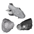
HREM has been widely used in multiple disciplines and can be applied to a wide range of morphological studies. Imaging denser samples up to 25mm down to below 1mm with voxel sizes down to 1 micron. Most commonly, samples in the range of 1-12 mm are imaged for detailed structural analysis.

Mouse Embryos
HREM has been widely used to image full mouse embryos across all stages of development.
-
Full 3D reconstruction of whole mouse embryos in high detail
-
Track developmental abnormalities across gestational stages
-
Quantify organ formation and spatial relationships

Small Animal Organs
Capture kidney, liver, placenta, lung and other small animal organs in full 3D from under 1mm up to 20mm, with consistent micron-scale section thickness for quantitative morphometric analysis.

Mouse Hearts
HREM has been used extensively for mouse heart imaging at stages E9.5 and above.
-
Visualise intricate 3D cardiac structures and vasculature
-
Study congenital heart defects (CHD) and structural remodelling
-
Ideal for phenotyping mutant mouse lines
Chick Embryos & Similar
-
Large scale imaging of early vertebrate development
-
Assess segmentation, heart looping and organogenesis

Plant Samples & Seeds
3D imaging of seed and root structures at micron resolution without sectioning artefacts.
-
3D imaging of seed structures and root development
-
Image vascular bundles, meristems and embryogenesis
-
Ideal for agricultural trait research and seed phenotyping
Flexibility for a Range of Samples
High Resolution Episcopic Microscopy is less impacted by optical penetration depth than other volumetric techniques, as imaging is performed directly on the resin block surface. This produces reliable three-dimensional imaging of structures including bone, calcified structures and pigmented tissue with complex samples with mixed density producing high contrast.
HREM supports imaging across a wide range of biological samples, including mixed density tissues within a sample.

Bone & calcified structures
Dense tissue imaged with consistent contrast, no clearing related artefacts or penetration limits.
Mixed density samples
Soft and hard tissue in the same block with no trade-off in contrast quality across the sample.
Pigmented tissue
No optical interference contrast is consistent regardless of pigmentation.
Intact whole samples
Up to 25mm in a single acquisition. Larger samples accommodated with scanning configurations.

High-resolution stitched Optical HREM Ultra image of a mouse section, revealing fine structural detail across the tissue.
High Resolution for Various Tissue and Sample Types
HREM typically operates within the voxel size range between 1-8 microns, with section thickness defining the Z resolution, enabling accurate 3D measurements and reliable structural reconstruction.
At these resolutions, fine structures can be visualised and quantified/segmented such as small vessels, branching networks, nerve structures and tissue boundaries. Unlike conventional imaging approaches where resolution can decrease with sample size, HREM maintains resolution with scanning configurations.
-
1-8 Micron voxel resolution for detailed volumetric imaging
-
Defined axial resolution via physical section thickness
-
Scanning configurations for extended field of view at maintained resolution
Configurable to your requirements
XY
SCANNING
Scanning stages
Extend field of view across larger samples. Resolution no longer limited by sample size.
EF
FOCUS
Electronic focus module
Adjust focus across the full block face with ease from the dedicated software application, for multiple samples or simply ease of use.
SI
ILLUMINATION
Structured illumination
Improves fluorescence contrast and rejects out-of-focus background for targeted channel imaging.
Det.
SESNORS
Detector options
Choice of detectors from low-noise sensors to high-resolution single-shot sensors, matched to your resolution and sensitivity needs.
4×
BLOCKS/RUN
Multiple samples
Image up to 4 blocks per session. Increases throughput for cohort phenotyping and knockout screening.
↗
CUSTOM
Custom configurations
Specific requirement? We work with you on optics, blade types, illumination, and software to meet your exact imaging needs.
Every HREM system can be upgraded or configured to match your application across resolution, throughput, illumination, and sample handling. Options shown with system compatibility.

Stitched Optical HREM 3x2
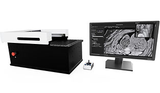
Optical HREM Systems
We offer two main Optical HREM system solutions, available worldwide, offering flexibility in budget and in capture. Each system can achieve highly detailed 3D image stacks, while allowing the systems to be accessible to more.

OHREM Micro
Micro OHREM Systems offer simple imaging of samples up to 25mm in FOV, featuring single shot optics.
Highlights
-
25 × 20 × 20mm field of view
-
Single-shot optics
-
Compact form factor
-
Optional scanning and focus module
.jpg)
OHREM Ultra
Ultra OHREM systems offer flexible imaging with XY scanning stages with various illumination and optical setups.
Highlights
-
Larger field of view with scanning
-
Structured illumination option
-
Multi-sample up to 4 blocks/run
-
Electronic focus module
Not sure which fits your work?
CHOOSE MICRO IF...
-
Samples are consistently under 20mm
-
Single-sample runs suit your workflow
-
Lab space or budget is a consideration
CHOOSE ULTRA IF...
-
Scanning or larger field of view needed
-
3+ Fluorescence channels are required
-
High throughput or multi-sample runs

Find out more
Browse our HREM products, application examples and imaging data or get in touch to discuss your specific sample.
Frequently asked questions
What sample types are suitable for High-resolution episcopic microscopy
Common examples of samples used in High-Resolution Episcopic Microscopy (HREM) include embryos, small organs, tissue blocks, plant material and other dense or opaque specimens. It works well for samples that are difficult to image using confocal or light-sheet microscopy, but can also complement these imaging methods.
What resolution can HREM acheive?
High-Resolution Episcopic Microscopy (HREM) resolution is typically between 1–8 µm voxel size, and the X–Y resolution is often higher than the vertical (Z) resolution.
What are the advantages of HREM over trafitional sectioning methods?
High-Resolution Episcopic Microscopy (HREM) eliminates the distortion and alignment issues common in manual sectioning. Each slice is imaged directly at the block face, so the resulting dataset is perfectly aligned and high in contrast. This produces cleaner 3D reconstructions, reduces manual handling, and speeds up the workflow compared to traditional sectioning methods.
How long does a typical HREM imaging session take?
HREM samples can be imaged at 6–7 seconds per section, though this varies with section thickness and sample size.
Approximate imaging speed per millimetre
-
1 µm → 1000 sections → ~1 hr 57 mins
-
2 µm → 500 sections → ~58 mins
-
3 µm → 333 sections → ~39 mins
-
4 µm → 250 sections → ~29 mins
-
5 µm → 200 sections → ~23 mins
-
Real-world example timings
-
Embryonic mouse heart (E14–E16, ~1.5–2.0 mm deep)
-
1 µm: 2.5–3.3 hrs
-
2 µm: 1.3–1.9 hrs
-
3 µm: 1.0–1.3 hrs
-
-
E10–E11 mouse embryo (small, ~4–6 mm deep)
-
1 µm: 6.7–11.7 hrs
-
2 µm: 3.3–7.0 hrs
-
3 µm: 2.2–3.9 hrs
-
-
E14–E15 mouse embryo (larger, ~8–10 mm deep)
-
1 µm: 13–19 hrs
-
2 µm: 6.7–9.7 hrs
-
3 µm: 4.4–6.5 hrs
-
What sample preparation is required for High-resolution episcopic microscopy?
HREM requires resin-embedding of the sample so it can be cut cleanly during imaging. Samples are fixed, dehydrated, infiltrated with resin and then polymerised before imaging. This preparation creates a stable block that allows consistent sectioning and produces clear, high-contrast images at each step.
What are the limitations of high-resolution episcopic microscopy?
HREM requires resin embedding and physical sectioning, so it is a destructive technique and cannot be used on live samples. It also cannot directly capture fluorescence during imaging, though markers are possible such as Lac-Z, and dataset sizes can be large. Sample preparation and resin processing take time, and highly calcified or very large samples may need additional preparation.
Do you supply High-resolution episcopic microscopy systems worldwide?
Yes. We supply High-Resolution Episcopic Microscopy (HREM) systems worldwide and provide remote support, installation guidance and ongoing servicing. Systems can be customised to your application, and we offer international shipping and technical assistance for laboratories outside the UK.
What support or training do you offer for High-resolution episcopic microscopy?
We provide ongoing support after installation, including remote assistance, software help, troubleshooting and guidance with imaging workflows. Training can be delivered on system operation, sample preparation and data handling to ensure your team is confident using the HREM system.
Contact our High-Resolution Episcopic Microscopy (HREM) Experts
Want to know more about HREM, ask for a quote or get questions answered. Contact us and we can help answer all your questions.
Phone:
+44(0) 1462633500
Email:
hello@indigo-scientific.co.uk